
When you are dealing with an injury or persistent musculoskeletal pain, one of the most common questions patients have is, “Do I need imaging?”
With so many different imaging options available today, it can be difficult to understand when an X-ray, CT scan, MRI, or diagnostic ultrasound is actually necessary. While imaging can provide valuable information that we cannot obtain from a physical examination alone, more imaging does not always mean better care.
The goal is to use the right imaging study at the right time to answer the right clinical question.
Why Is Advanced Imaging Helpful?
It Allows Us to See Structures That Cannot Be Directly Evaluated on a Physical Examination
A physical examination can tell us a lot about how a patient moves, where they hurt, what movements reproduce their symptoms, their strength, and whether there are neurological deficits. However, there are limitations to what we can determine through a physical examination alone.
We cannot directly visualize the condition of a tendon, ligament, muscle, disc, cartilage, or bone simply by examining how someone moves or where they feel pain. This is where imaging can provide valuable additional information by allowing us to visualize structures beneath the surface that cannot be directly assessed during a physical examination.
Imaging Can Identify Structural Injuries and Help Guide the Plan of Care
Imaging can help identify structural abnormalities such as fractures, tendon or ligament injuries, muscle tears, joint abnormalities, degenerative changes, disc pathology, and other structural changes. It can also help determine the extent of an injury, which structures are involved, and whether multiple structures may be affected. Because different tissues and structures can have different healing timelines, this information can help establish a more realistic prognosis and guide the plan of care.1
| Tissue | Healing Time | |
|---|---|---|
| Muscle | Exercise Induced | 0 – 3 days |
| Grade 1 | 1 – 4 weeks | |
| Grade 2 | 3 – 12 weeks | |
| Grade 3 | 1 – 6 months | |
| Tendon | Tendonitis | 3 – 7 weeks |
| Tendonosis | 3 – 6 months | |
| Ligament Sprain | Grade 1 | 2 – 8 weeks |
| Grade 2 | 2 – 6 months | |
| Grade 3 | 6 – 12 months | |
| Meniscus / Labrum | 3 – 12 months | |
| Fracture | 6 – 8 weeks | |
In some cases, imaging confirms that conservative treatment is appropriate. In others, it may identify a condition that requires a different level of care, such as referral to an orthopedic specialist, surgical consultation, additional diagnostic testing, different rehabilitation recommendations, or more appropriate activity restrictions.
When Is Imaging Appropriate?
Imaging isn’t necessary for every injury or episode of musculoskeletal pain. However, there are certain situations where imaging can provide information that may change the diagnosis, prognosis, or plan of care.2
Imaging may be appropriate following a significant traumatic event, particularly when there is concern for a fracture or significant soft-tissue injury. It may also be indicated when red-flag symptoms are present, including:
- Severe, sudden, or progressively worsening neurological symptoms
- Bowel or bladder changes
- Saddle paresthesia
- Unexplained weight loss
- Constant, unexplained night pain
- A personal history of cancer
Imaging may also be appropriate when a condition fails to improve with conservative care or progressively worsens. When a patient is not recovering as expected, imaging can help identify an underlying structural problem that may not have been apparent on physical examination, or determine whether the original diagnosis or treatment plan should be reconsidered.
When Is Imaging Not Appropriate?
Just because imaging is available does not mean it will provide useful information. Unnecessary imaging can add cost, expose patients to radiation when applicable, and identify findings that may be unrelated to their symptoms.2
A simple question to consider before obtaining an image is:
If the results are positive or negative, will they change my treatment plan or the patient’s next step in care? If the answer is no, imaging may not provide meaningful clinical value.
The goal isn’t to obtain the most advanced imaging available. The goal is to obtain the right imaging when it can meaningfully change the patient’s care.
What Imaging Options Are Available?
There are several types of diagnostic imaging, and each has its own strengths and limitations. The best choice depends on the structures we need to evaluate and the clinical question we are trying to answer.
X-Ray
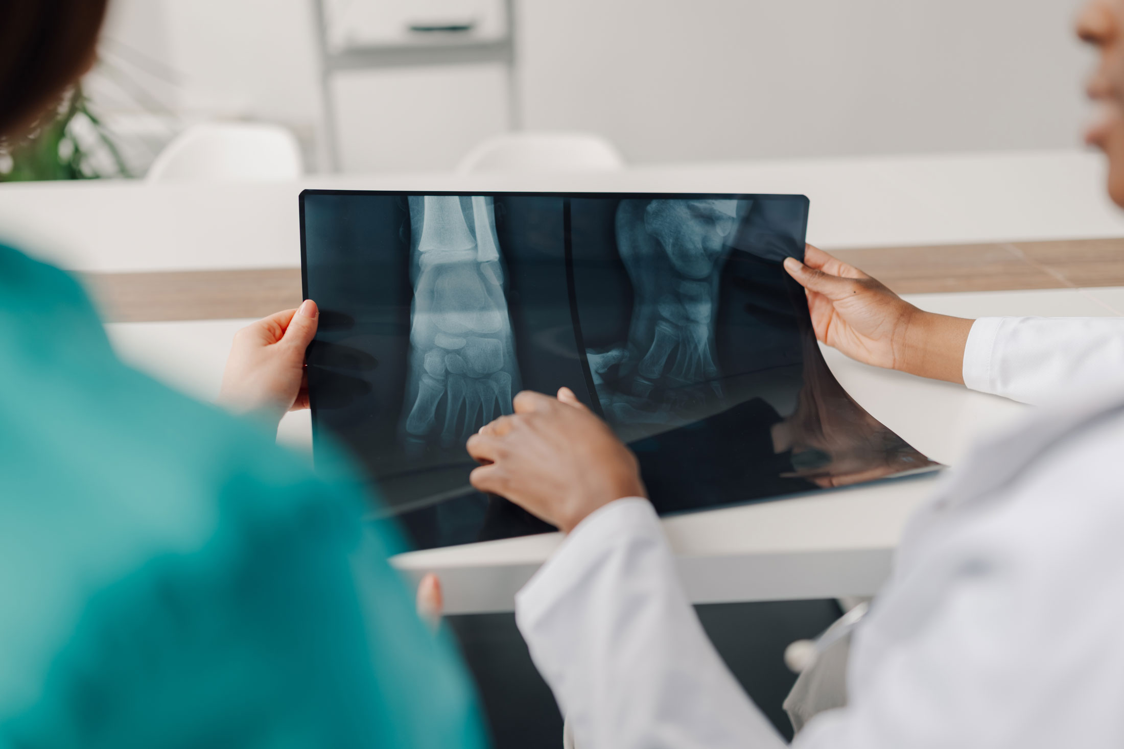
X-rays are one of the most common forms of medical imaging and are particularly useful for evaluating bone and alignment.3
What Are X-Rays Good At?
- Identifying fractures
- Evaluating bone
- Assessing alignment
- Evaluating certain joint changes
- Identifying certain degenerative changes
X-rays are also fast, relatively inexpensive, and widely accessible. They do use ionizing radiation, but the radiation exposure from a conventional X-ray is relatively low and varies depending on the body part being examined.
One of the main limitations of X-rays is their ability to visualize soft tissue. Tendons, ligaments, muscles, discs, and other soft tissues are difficult to evaluate with conventional X-rays.
CT Scan
A CT scan, or computed tomography scan, uses X-rays to create detailed cross-sectional images of the body. CT scans are particularly useful for evaluating bone and deeper or more complex structures.4
What Are CT Scans Good At?
- Evaluating complex fractures
- Evaluating the skull and spine
- Evaluating the chest, lungs, and abdomen
- Identifying certain calcifications and stones, such as kidney stones and gallstones
One of the main advantages of CT is that it is relatively fast compared with MRI, making it particularly useful in emergency situations and following significant trauma.
The biggest consideration with CT is that it uses ionizing radiation. The radiation exposure is substantially greater than that of a conventional X-ray, although the actual dose varies depending on the type of CT examination.
Because ionizing radiation carries a potential long-term cancer risk, CT should be used when the expected diagnostic benefit outweighs the potential risk.5 This does not mean CT scans are inherently dangerous or should be avoided. When medically necessary, the information they provide can be extremely valuable.
Diagnostic Ultrasound
Diagnostic ultrasound uses high-frequency sound waves to create images of structures within the body. It is particularly useful for evaluating superficial soft-tissue structures.6
What Is Diagnostic Ultrasound Good At?
- Evaluating tendons
- Evaluating muscles
- Evaluating ligaments
- Evaluating certain joints
- Evaluating other superficial soft tissues
- Time efficient
One of the biggest advantages of ultrasound is its ability to provide real-time, dynamic visualization. Unlike X-ray, CT, and MRI, ultrasound allows us to watch certain structures while the patient moves. This can be particularly useful when symptoms occur during a specific movement or when evaluating how a tendon or other structure behaves dynamically.
Ultrasound also does not use ionizing radiation, which makes it safe for almost every type of patient.
However, ultrasound does have limitations. It is not ideal for evaluating very deep structures or structures obscured by bone or air. For example, ultrasound has limited ability to evaluate structures deep within the spine.
At M3 Chiropractic & Sports Medicine, diagnostic ultrasound is one of the specialized tools we have available to help evaluate certain musculoskeletal injuries and conditions.
MRI
Magnetic resonance imaging, or MRI, uses a powerful magnetic field and radiofrequency energy to create detailed images of the body’s internal structures. Unlike X-ray and CT, MRI does not use ionizing radiation and is particularly useful for evaluating soft tissues and deeper structures.7
What Is MRI Good At?
- Evaluating muscles
- Evaluating tendons and ligaments
- Evaluating joints
- Evaluating discs and nerves
- Evaluating bone marrow
- Evaluating other soft tissues
MRI is often used when a more detailed evaluation is needed for structures that cannot be adequately evaluated with an X-ray.
MRI does have some limitations. It is generally more expensive and time-consuming than X-ray or ultrasound, and patients must remain still for an extended period of time. The enclosed space can also be challenging for patients with claustrophobia. Unlike diagnostic ultrasound, MRI provides primarily static images rather than real-time visualization during movement.
So Which Imaging Test Is Best?
There isn’t one imaging modality that is universally better than the others. Each type of imaging has different strengths and is best suited for different clinical questions.
- X-ray is excellent for evaluating bone, fractures, alignment, and certain joint changes.
- CT provides detailed cross-sectional images and is particularly useful for bone and complex anatomy.
- Diagnostic ultrasound is excellent for many superficial soft tissues and provides the unique advantage of real-time, dynamic visualization.
- MRI provides excellent detail of many soft tissues and deeper structures without using ionizing radiation.
The important question isn’t, “What is the most advanced imaging test available?” but rather, “What imaging study will best answer the clinical question we are asking?”
The Bottom Line
Imaging isn’t necessary for every episode of musculoskeletal pain. In many cases, a thorough history and physical examination provide enough information to make a diagnosis and begin appropriate treatment. When imaging is appropriate, the goal is not simply to obtain the most advanced test available, but to choose the modality that will provide the information needed to guide care.
Ultimately, imaging should complement a thorough history and physical examination, not replace them. The image is one piece of the clinical picture—not the entire picture. A good rule of thumb for providers to follow is to treat the patient and their clinical presentation, not simply the image.
If you’re dealing with an injury or persistent musculoskeletal pain and aren’t sure whether imaging may be appropriate, schedule an evaluation with M3 Chiropractic & Sports Medicine. We can help determine whether additional imaging is necessary and, if so, which type may provide the most useful information to guide your care.
References
- Kirkby Shaw K, Alvarez L, Foster SA, Tomlinson JE, Shaw AJ, Pozzi A. Fundamental principles of rehabilitation and musculoskeletal tissue healing. Vet Surg. 2020 Jan;49(1):22-32. doi: 10.1111/vsu.13270. Epub 2019 Jul 4. Erratum in: Vet Surg. 2025 May;54(4):647. doi: 10.1111/vsu.14253. PMID: 31271225; PMCID: PMC6973127.
- Expert Panel on Neurological Imaging; Hutchins TA, Peckham M, Shah LM, Parsons MS, Agarwal V, Boulter DJ, Burns J, Cassidy RC, Davis MA, Holly LT, Hunt CH, Khan MA, Moritani T, Ortiz AO, O’Toole JE, Powers WJ, Promes SB, Reitman C, Shah VN, Singh S, Timpone VM, Corey AS. ACR Appropriateness Criteria® Low Back Pain: 2021 Update. J Am Coll Radiol. 2021 Nov;18(11S):S361-S379. doi: 10.1016/j.jacr.2021.08.002. PMID: 34794594.
- Tafti D, Maani CV. X-ray Production. [Updated 2023 Jul 31]. In: StatPearls [Internet]. Treasure Island (FL): StatPearls Publishing; 2026 Jan-. Available from: https://www.ncbi.nlm.nih.gov/books/NBK537046/
- Singh V, Sandean DP. Computed Tomography Safety. [Updated 2023 Jan 2]. In: StatPearls [Internet]. Treasure Island (FL): StatPearls Publishing; 2026 Jan-. Available from: https://www.ncbi.nlm.nih.gov/books/NBK567800/
- Smith-Bindman R, Chu PW, Azman Firdaus H, Stewart C, Malekhedayat M, Alber S, Bolch WE, Mahendra M, Berrington de González A, Miglioretti DL. Projected Lifetime Cancer Risks From Current Computed Tomography Imaging. JAMA Intern Med. 2025 Jun 1;185(6):710-719. doi: 10.1001/jamainternmed.2025.0505. Erratum in: JAMA Intern Med. 2025 Jun 1;185(6):747. doi: 10.1001/jamainternmed.2025.2355. PMID: 40227719; PMCID: PMC11997853.
- Wagai T. Studies on the foundation and development of diagnostic ultrasound. Proc Jpn Acad Ser B Phys Biol Sci. 2007 Dec;83(8):256-65. doi: 10.2183/pjab/83.256. PMID: 24367150; PMCID: PMC3859294.
- Al-Naser YA, Tafti D. Magnetic Resonance Imaging (MRI) Safety and Biological Effects. [Updated 2026 Jun 19]. In: StatPearls [Internet]. Treasure Island (FL): StatPearls Publishing; 2026 Jan-. Available from: https://www.ncbi.nlm.nih.gov/books/NBK604477/